Diagnosing Diverticulitis

When diagnosing diverticulitis, the first thing that doctors usually do is carry out a blood test followed by other tests such as CT scans, X-rays and even ultrasounds.

Full Blood Count

The full blood count can tell doctors a lot about your current health. The number of red blood cells can tell doctors if you’re anaemic and perhaps losing blood. But one of the most important markers in a blood test when trying to check for diverticulitis is the white blood cell count.

White blood cells are a part of the immune system and are responsible for fighting diseases and illnesses. When a pathogen (harmful bacteria or virus) enters the body, it is identified by the immune system which then increases the number of white blood cells. An amount of white blood cells higher than the normal range could be considered to be a sign of infection. However, it is possible to have diverticulitis but not have a measurable increase in white blood cells. So a normal blood result doesn’t necessarily mean that you don’t have an infection or inflammation in the bowel.

CRP (C-Reative Protein)

CRP is known as an inflammation marker – it is a protein released by human tissues when they are inflamed. The higher the amount of CRP in the blood, the more inflammation there is. Having elevated CRP in addition to abdominal pain and other symptoms is a good indicator of diverticulitis. However, your doctor should also check carry out an abdominal examination too just in case the inflammation is coming from somewhere else.

An abdominal exam can help doctors find out if there is anything strange going on…

Abdominal Examination

It’s likely your doctor may carry our an abdominal examination. This is where the doctor will press on your abdomen to discover where the pain originates and if it is the bowel that is inflamed. It’s important to be very honest about the pain you are feeling at this point so that the doctor can rules out any other issues or sources of inflammation.
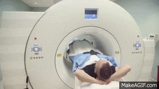
CT scanner…in you go!

CT scan

This is usually the most effective way to diagnose diverticulitis. A CT scan can be done with or without contrast, but contrast certainly helps when interpreting the results. The two main types of contrast are oral and intravenous (IV). The oral contrast is often a liquid mixed with water that you drink before the scan. It helps to highlight the digestive tract on your scan. The IV contrast is injected into a vein and reaches the tissues and helps them show up better on the scan. Some hospitals may choose to have their patients take both types of contrast. However, it is important than you let the doctor know of any allergies you have before you receive any contrast agents.

A CT scan is painless and just requires the patient to hold their breath for short periods. Let the technician or doctor performing the scan know if you suffer from asthma or COPD or any problem that makes you find it difficult to hold your breath.

After the scan, your results will not be ready immediately since they will need to be looked at by a doctor and a thorough report written. See my post on CT scans for more information.

X-rays

Some doctors may choose to send their patients for X-rays. This is to check for perforations in the bowel since X-rays can easily pick up excess gas in the abdomen that may be leaking out from the bowel. The results of an X-ray are much more immediate which is why this test might be chosen particularly if the patient is in a lot of pain or has a history of perforations and sepsis.

Ultrasound

Again, some doctors may want to send their patients for an ultrasound. This is very common during first time diagnoses when patients are doctors don’t know that the patient even has diverticular disease yet. An ultrasound may also show an abscess or cyst as a result of diverticulitis. It may also be able to detect excess air of material in the abdomen. However, it is not as clear as a CT scan, but the results are more immediate.

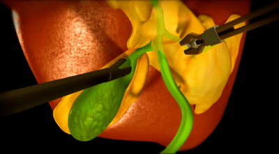
Why don’t doctors use colonoscopies to diagnose diverticulitis?

It’s very rare that a doctor would use a colonoscopy to diagnose diverticulitis because while a patient has an infection, it would be very painful and a little dangerous to insert an endoscope. There is a chance that the colonoscopy during an infection could make it even worse or could even cause damage to the colon.

Have you been diagnosed with diverticulitis? Which methods did the doctors use to diagnose you?

Gallbladder removal surgery – Cholescystectomy

Gallbladder removal or a cholescystectomy is a common surgery carried out by surgeons all over the world. The gallbladder is an organ located in our upper-right abdomen, just beneath the liver. It is the place where bile is stored. Bile is a yellow/green liquid produced by the liver when it breaks down substances in the body (including our old red blood cells).

The job of bile in the body is to emulsify fats – it does this by breaking up the fats and oils we consume into smaller droplets so that our body can digest it more easily. It also helps to neutralise the acidic chyme (food + stomach acid mixture) that leaves the stomach and enters the small intestine.

The reasons for a cholescystectomy include;

  • gallstones
  • gallbladder disease/infection / cholecystitis
  • non-functional gallbladder/gallbladder sludge
  • polyps

So what is the surgery like?

Well, these days, most cases are done laparoscopically. This means that it is done using several small incisions in the abdomen (for example in the navel, right side of the abdomen and possibly an incision in the centre of the upper-abdomen. Most patients have three or four incisions, but there are cases of people having less or more. The surgery is carried out under general anaesthetic. This means that you’ll be asleep and unaware of anything going on. Most patients are intubated (with a breathing tube) for this procedure, but are not usually catheterised. The surgery usually lasts between one and two hours. Depending on your hospital, some people may go home the same day and in others, they may be asked to stay in hospital overnight.

If you have an open surgery, the surgeon will make a large incision in the upper-right abdomen and take the gallbladder out from here. This is avoided where possible since it increases the length of time needed for recovery and hospitalisation.

During a laparascopic surgery, the surgeon will inflate the abdomen with gas. This helps provide more space in side to move around and perform the procedure. Then a camera will be inserted into one of the incisions so that the surgeon can see what he is doing. After this, various tools will be inserted into incisions to cut the gallbladder free. Medical titanium surgical clips will be placed to cut off tubes that connected the liver to the gallbladder. These metal clips will remain inside your body for the rest of your life.

Of course like any surgery, it does carry risks and there are complications that can arise. However, for this surgery, complications are rare and most patients deal with it very well.

What is the recovery period like?

Well, for me, it wasn’t too difficult. But everyone heals at different speeds and has a different experience without their gallbladder. Some people are back at work within a few days, and others may take a couple of months to get back to normal.

My advice is not to rush yourself. Recovery is an important process for our body and to recover well you need to rest and relax while trying to stay healthy. Your surgeon will give you some guidelines for aftercare and recovery, so it’s important to stick to those as much as your possibly can.

General guidelines include:

  • No lifting/carrying for 6 weeks – During the surgery the abdominal muscles are often cut. Heavy lifting, carrying or even straining on the toilet can cause these muscles to separate and increase your risk of a hernia.
  • No baths – whether your surgeon has used stitches or medical glue, it isn’t advisable to take a bath too early on. The water can soften the healing tissue or the glue and can increase the risk of infection. Taking a shower might be okay, but you should ask your surgeon if they recommend covering the incisions with waterproof dressings beforehand.
  • Check the incision sites regularly for redness, feeling warm to the touch and weeping. Clear fluid weeping from incisions may be normal, but if the fluid is yellow and opaque and/or you have any of the above symptoms, it may be worth mentioning to your surgeon.
  • Stick to a post-op gallbladder diet if given one.
  • Take regular, gentle exercise but DON’T overdo it!

To read about my gallbladder story click here! Want to share your story? Get in touch with me so that we can tell your story, too!

Hayley’s Gallbladder story

My gallbladder story is quite short really and uncomplicated in comparison to others. I’d never really experienced any problems with my gallbladder. Although, looking back, there was a time I had a sharp and horrendous pain in my upper-right abdomen in 2012 one day while at work. The pain was severe and my boss called an ambulance which came right away and injected me with a load of pain killers and muscle relaxants. I went home and slept it off and the pain was gone within a few hours so I never even went to a doctor about it. Maybe, that was an attack, maybe not.

Fast-forward to the beginning of February 2019. I was still being investigated after a complicated case of diverticulitis that resulted in the perforation of my bowel and a battle with sepsis. My constant lower-abdominal discomfort caused my gastroenterologist to send me for a full abdominal ultrasound. He wanted to rule out abscesses, cysts and adhesions so requested a scan of the whole abdomen.

Ultrasounds are no big deal. Totally painless and easy to do.

At the ultrasound, the doctor started snapping pictures of my organs and measuring dimensions, then, the moment he got a clear image of my gallbladder, he asked ‘Are you on the waiting list to get this removed?’, ‘No’ I replied, ‘Why do you ask?’ and he told me that there were a few small stones in there, but more importantly a polyp. I told him I had no idea and hadn’t had any symptoms of things not working well. Anyway, at the end of the ultrasound, he gave me all the printed photographs and a written report detailing what he had found and told me to go back to my gastroenterologist as soon as possible.

The gastroenterologist told me that it would need to be removed and set up a meeting with a surgeon for a few days later. He looked at my results, agreed that it needed to be removed because of the potential for the polyp because of its size to be a risk. He penciled me in for two weeks later to get it removed. In the mean time, he told me to eat a low fat diet and gave me a list of foods to avoid. (I’ll post about this soon, I promise)

Before I knew it, it was removal day. At 7:30am I hopped onto the trolley and was taken down to surgery. I watched the staff running around doing last-minute checks and preparation. It was fun chatting to the hospital porter who spoke pretty good English and was so smiley. I wasn’t particularly nervous or scared, I deal with hospital and needles etc pretty well and I’m not someone to get themselves into a state about a surgery. I’m sure I was mid-sentence joking with the surgeon and porter as I drifted off into a deep sleep and the surgery began.

In what seems like no time at all, I wake up in a different room with an IV in the opposite arm and a drain coming out of a hole in my upper-right abdomen. I look to my right and the porter is stood there smiling. I smile back and say ‘I need to sit up’, He immediately props the trolley up and hands me a little sick bowl….no, I say, I feel like I can’t breathe. So, he calls over a nurse who measures my oxygen levels and rushes off and comes back with a mask. I’m hooked up to oxygen and starting to catch my breath. Five or ten minutes later, the porter takes me back to the ward where my boyfriend was waiting to see me. He seemed worried, I asked him what the problem was and it was only then, that he told me that the surgery was expected to last maybe an hour and a half, but that I had been there for over four hours due to it being a tricky surgery. I hadn’t even noticed what the time was.

I expected to be in major pain the rest of the day, but other than a horrible discomfort in my back and shoulders, I had no pain at all. I refused dinner when the nurse brought it. I wasn’t hungry at all and the noodle soup did NOT look appetizing at all. I was alone now because my boyfriend had left for work. I forgot I had the drain and as I got up out of bed and went to the bathroom, I must have moved the drain a little and it was a bit sore. After the effort of that, I went back to bed and was in a little pain. The nurse came to check on me and when I told her it was sore, she injected something into my IV and the pain subsided and I drifted off again.

I woke up the next morning feeling quite chipper. I still had some discomfort in my back and chest from the gas, but was keen to get up and about. I got up had a wash, brushed my teeth and did my hair. Then I got changed. When the nurse offered pain relief via the IV, I declined since I didn’t have any pain except for the gas pains. I got up and walked around the ward and the catering team brought me a chamomile tea which I sipped at.

My surgeon came in to speak to me and told me not to lift heavy things or to strain when going to the bathroom due to the risk of getting a hernia. I was a little worried since I had pushed a bit going to the toilet in the morning, but it was okay, the stitches had held. He removed the drain and replaced my dressings. Before telling me to take almost 3 weeks off work and discharging me from the hospital. He told me to clean my wounds and replace the dressings each day. He also  advised me to take a paracetamol if I was uncomfortable, but to contact him if I had severe pain, or jaundice.

I didn’t even need the paracetamol. Recovery wasn’t too difficult, but I stuck to the post-op gallbladder diet that he gave me. (I promise I’ll write about this, too) The one thing I had a problem with, was laying down. The stitches were tight and I couldn’t lay down or get up, so for the first week, I slept sitting up on the couch. It wasn’t a problem.

Sleeping on the couch wasn’t so bad!

I used glycerine suppositories everyday to keep me going to the bathroom without effort. As the days passed, I found doing things even easier and as time passed I found myself able to tolerate more foods. The surgeon checked up on me regularly via telephone and asked me to send him pictures of the healing incisions on Viber. They were all doing great accept for my belly button which was weeping and a little sore and hot to the touch. Although, the surgical incision in my belly button did become a little infected and I had to pack it and take antibiotics, but within a week, it was much better and I could go to have the stitches removed. I had to be careful to stay near a toilet after trying a new food though because a few times in those first couple of months, I experienced dumping syndrome and had to make a bee line for the bathroom.

Post-op, life isn’t much different for me. I sometimes still get dumping syndrome, particularly if I eat something too fatty. Otherwise, I don’t have any real symptoms…just like before my op. However, it’s nice to know that the polyp was removed and won’t be causing any trouble in the future.

So, if you’re having your gallbladder removed. Don’t worry. It isn’t as bad as you might think. If you are suffering with a lot of anxiety, tell your doctor. There are things they can do to help you relax and to reassure you about the surgery. Of course, everyone’s gallbladder story is a little different. But, this is mine and from my experience, I feel it wasn’t a bad or scary experience. But, it is so important to take time out to recover.

My advice is to not push yourself or overdo it during your recovery – you may regret it. Also, don’t expect to feel back to normal immediately. The human body is amazing, but it certainly needs to be given the time to heal after surgery.

Diverticular Disease Tracker

Keeping your food, symptom and stool diary

Keeping a diary isn’t always easy, but…it can come in very handy, especially if you are still learning to manage a health condition. If you are someone that already keeps a daily diary or journal, it may be easy to add this information in to your entries. However, if you’re not someone used to keeping a diary, then you can find other ways to do it, for example, you can use the downloadable attachment here, and complete that each day before filing it away, or you can create your own version that applies more specifically for you. Not only will this help you identify trigger foods or patterns in your symptoms but it could also be a great tool to help your doctors monitor and manage your health.

NOTES:

Be honest! Write what you really ate and drank….snacks, junk and alcohol included…you need to see the whole picture, exactly as it is.

Take it to doctor’s appointments with you even if you don’t need to use it. You never know when the info could come in handy.

If you have a bullet journal, you can get really creative with this. I’ve given you guys a free downloadable and printable version of what I use. Now, unfortunately you do need to print it to get the best use out of it. I would recommend filing them each day in a binder to keep a continuous diary. How much additional information you record is totally up to you. However women may benefit also by tracking their menstrual cycle, too.

DD Tracker Printable

I’ve put together a little Diverticular Disease/Diverticulosis Tracker. You can use this to monitor what you eat, how you feel and even keep tabs on your bowel movements. I hope this proves to be helpful. The file even has it’s very own instructions that show you exactly how to use it. Just click on the download button below for FREE and you can print it out right away.
Support the creator and buy her a coffee by making a donation here: paypal.me/haylaki

Food Diary: The food sections allow space for you to write about what you ate and drank for the day.

Water Intake: You can cross out or tick the glass of water to show how much you drink each day.

Bowel Movements: The stool section provides boxes for you to keep a record of your bowel movements. There is space to write a time and or comment and a number that corresponds to the Bristol stool chart.

Symptoms/other notes: here you can mention any other feelings or symptoms you experience even if you feel they aren’t relevant. For example, headache, joint pain, nausea, delicate mood, etc. or even record that you took pain medication or anything that you don’t usually take. In addition, you could note anything else there you wish.

Medication: It’s always worth making a note of any medication you had to take, whether it seems related or not. But taking medication such as painkillers or antacids is a good way to keep a record of how often your symptoms bother you enough to reach for the medicine cupboard.

Tracker Previews

Preview of the DD Tracker
Instuctions and examples

Let me know what you think and even share with me what you record in your diary to help you…do you do anything differently?